Sign up for the Design News Daily newsletter.
New AI Allows Physicians to Obtain Better Images of the Heart
The success of the approach was chronicled in a new article in the scientific journal Circulation.
MDDI Staff
August 31, 2021
1 Min Read

Yevhenii - stock.adobe.com
Researchers from University of Virginia Health System have developed an artificial intelligence technology for heart imaging – dubbed the Virtual Native Enhancement (VNE). The researchers said the AI technology could improve care for patients allowing doctors to examine their hearts for scar tissue while eliminating the need for contrast injections required for traditional cardiovascular magnetic resonance imaging (CMR).
The success of the approach was chronicled in a new article in the scientific journal Circulation.
The new VNE technology will allow doctors to image the heart more often and more quickly, the researchers say. It also may help doctors detect subtle changes in the heart earlier, though more testing is needed to confirm that.
The technology also would benefit patients who are allergic to the contrast agent injected for CMR, as well as patients with severely failing kidneys, a group that avoids the use of the agent.
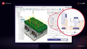
The new approach works by using AI to enhance "T1-maps" of the heart tissue created by magnetic resonance imaging (MRI). These maps are combined with enhanced MRI "cines," which are like movies of moving tissue - in this case, the beating heart. Overlaying the two types of images creates the artificial VNE image
Based on these inputs, the technology can produce something virtually identical to the traditional contrast-enhanced CMR heart scans doctors are accustomed to reading - only better, the researchers conclude.
While the new research examined VNE's potential in patients with hypertrophic cardiomyopathy, the technology's creators envision it being used for many other heart conditions as well.
About the Author(s)
You May Also Like
Editors' Choice


Jun 4 - Jun 6, 2024
Jun 4 - Jun 6, 2024
Innovation in automation starts here. Discover and collaborate on automation solutions that are revolutionizing the entire production lifecycle — from design to production to market — and sharpen your competitive edge. ATX South is part of IME South, a six-in-one expo offering the latest insights & solutions spanning medtech, packaging, automation, plastics, design, & processing.
Register Now