

Sign up for the Design News Daily newsletter.
Researchers have created a new way to fill in cranium bones by using 3D printing technology.
Elizabeth Montalbano
April 4, 2018
5 Min Read

Medical applications have benefited greatly from 3D printing technology. Researchers continue to innovate, offering a continual stream of 3D printing advancements in medical treatment. Recently researchers demonstrated a new method that paves the way for live 3D printing of regenerative bone materials inside the human cranium designed to repair severe traumatic injuries—the first of its kind.
|
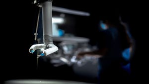
An image shows bone grafts used to fill large volume cavities to repair cranium defects. Dr. Venu Varanasi from the University of Texas and Texas A&M University College of Dentistry led a team that developed a technique to use 3D-printed scaffolds to replace bones in severe cranium injuries in a similar way to how cavities are filled at the dentist. (Source: U.S. patent application US20170143831A1) |
Led by Dr. Venu Varanasi from the University of Texas and Texas A&M University College of Dentistry, the research showed how the current use of plastic or metal implants to heal cranial injuries can be replaced with an approach akin to how dentists provide fillings for decayed teeth, he said.
"Our goal is to heal the defect or fracture site rapidly, as if nothing ever happened,” Dr. Varanasi said. "We want to develop these methods and materials so that someday we can treat certain types of bone defects like they are dental fillings. Principally, these become out-patient procedures where the patient goes home to heal with the support of their loved ones and with reduced medical expenses owed to extended hospital stays."
Creating Printed Scaffolds for Bone Healing
Specifically, Varanasi and his team—who presented their findings recently at 47th Annual Meeting of the American Association for Dental Research in Florida—demonstrated in a lab the possibility of 3D printing bone-scaffold substitutes during surgery that conform to the dimensions of a defect site.
Researchers tested several nanobiosilica-based 3D scaffolds with 3D-printing properties to “potentially improve implantability and rapid bone healing capability,” Varanasi explained. They using a human periosteum cell culture model and a rat cranial defect animal model to demonstrate the scaffold and live 3D printing method for “potential clinical translation,” he said.
Researchers prepared the biosilica-biopolymer scaffold by mixing Laponite with methacrylated gelatin. They used sucrose to increase viscosity and reduce gelation of the printing ink and IRGACURE 2529 as a crosslinking agent.
During printing, ultra-violet light at the tip of the printer nozzle initiated crosslinking, and scaffolds were in-situ 3D printed directly into calvaria bone defects using varied Laponite concentration. This determined optimal bone density and chemical structure, researchers said.
Fixing Bones During an Office Visit
The technique—which could be done in an outpatient way, much like a person goes to a dentist to fill a cavity—would be far less invasive and more conducive to rapid healing than how cranium defects are repaired now, researchers said.
Currently, the implants used must be customized for fit and therefore take time to develop. Once implanted, it also takes time for the existing bone to accept the implant and heal properly, sometimes requiring multiple surgeries during the process.
Not only can the 3D-printed scaffold researchers developed support rapid and complementary repair, researchers also can use the microarchitecture and materials chemistry of the scaffold to enhance tissue regeneration and growth to hasten the healing process, Varanasi said.
Quicker Healing with 3D Print Method
In lab tests, researchers fabricated scaffolds into a mesh design with dimensions matching that of formed defects. After four weeks, they extracted cranial bone samples and evaluated them by micro-CT.
Results showed that nearly 55 percent of the bone defect was healed after four weeks for higher Lp- rich-MAG scaffolds versus lower Lp-containing MAG scaffolds, according to the team. In contrast, empty control defects only had 11 percent of the defect filled with bone after four weeks.
Moreover, histological staining showed that the scaffolds recruited cells into their structure to regenerate the intra-bony layers needed to initiate the healing process, researchers said.
Overall, tests demonstrated that 3D in-situ printing of bone-regenerating scaffolds improve the delivery of regenerative and reconstructive biomedical devices for the proper and rapid healing of bone fractures, Varanasi said.
“The method allows for the absorption of blood and growth factors into the scaffold as it is being constructed within the defect,” he said. “This provides an advantage in that cells from within the initial hematoma become incorporated into the scaffold structure, thus giving the operator flexibility to use the printed scaffold as a structural support that stimulates healing.”
Varanasi and the team have filed a patent for their technique with the U.S. Patent and Trademark Office.
READ MORE ARTICLES ON 3D Printing:
Researchers will continue to pursue the work further as part of the Bone-Muscle Group at the University of Texas at Arlington College of Nursing and Health Innovation and in collaboration with the University of Texas at Arlington College of Engineering, Texas A&M University College of Engineering, and College of Dentistry, they said.
Elizabeth Montalbano is a freelance writer who has written about technology and culture for 20 years. She has lived and worked as a professional journalist in Phoenix, San Francisco and New York City. In her free time she enjoys surfing, traveling, music, yoga and cooking. She currently resides in a village on the southwest coast of Portugal.
As the Internet of Things (IoT) pushes automation to new heights, people will perform fewer and fewer “simple tasks.” Does that mean the demand for highly technical employees will increase as the need for less-technical employees decreases? What will be the immediate and long-term effects on the overall job market? What about our privacy and is the IoT secure? These are loaded questions, but ones that are asked often. Cees Links, wireless pioneer, entrepreneur, and general manager of the Wireless Connectivity business unit in Qorvo, will address these questions, as well as expectations for IoT’s impact on society, in this ESC Boston 2018 keynote presentation, Thursday, April 19, at 1 pm. Use the Code DESIGNNEWS to save 20% when you register for the two-day conference today! |
About the Author(s)
You May Also Like
Editors' Choice

Jun 4 - Jun 6, 2024
Jun 4 - Jun 6, 2024
Innovation in automation starts here. Discover and collaborate on automation solutions that are revolutionizing the entire production lifecycle — from design to production to market — and sharpen your competitive edge. ATX South is part of IME South, a six-in-one expo offering the latest insights & solutions spanning medtech, packaging, automation, plastics, design, & processing.
Register Now